Forearm (Radius & Ulna) Fractures
What is a Forearm Fracture?
The forearm is made up of two bones, the radius and the ulna. These can either be broken separately or together. This can occur in patients of all ages from a variety of traumatic causes. The bone can break in many different ways and can range in severity. If only one bone breaks a dislocation of the wrist or elbow is often present. Sometimes the bone breaks and sticks out through the skin. This is called an open fracture and needs urgent surgery. Each fracture type requires individual treatment so it is important for your doctor to diagnose and treat your injury appropriately.
What does the Forearm do?
The function of the forearm is to connect the elbow to the wrist. The shape of the forearm bones is important because it allows for rotation. This is what makes it possible to turn your palm up and down. Alignment of the forearm bones also keep the wrist and elbow joints moving correctly. Poor position can affect the ability to bend of straighten the elbow or wrist.
Diagnosis and Examination
Physical examination is important in the evaluation of these injuries. Important blood vessels and nerves lie near the bones and can be injured when they break. The doctor will also look for any open wounds over the injury. Sometimes the bone pushes against the skin causing it to “tent.” Too much pressure can result in the bone coming through the skin.
X-rays are used to evaluate the location, type, and severity of the broken bone. This helps doctors and patients make an informed decision on treatment. Often three or more x-rays are taken to show the injury pattern. X-rays of the wrist and elbow are also indicated to diagnose any associate dislocation or injury.
Non-Surgical Treatment
Many forearm fractures in children do not require surgery and can be treated in a cast. In adults, if only one bone is broken and it is well aligned, non-operative care can be done. However, most adult forearm fractures require surgery for good outcomes
If non-operative care is chosen, regular follow-up care for a physical exam and x-rays is important to ensure that the fracture stays in good position and heals appropriately. Cutting down or quitting smoking and tight blood sugar control if you are a diabetic is important for the healing process. If the bones lose their alignment or slip, surgery may be required.
Forearm fractures treated non-operatively are usually casted for six weeks. The cast is then removed, patients are given a removable wrist brace and physical therapy is started to help patients regain their range of motion.
Surgical Treatment
Surgeons recommend an operation to fix the broken forearm if it is broken into many pieces, if the bones are far apart, if both bones are broken, if the bone sticks out through the skin, or if the nerves or blood vessels are injured. If the bone has made a hole in the skin or is sticking out, urgent surgery is required.
Most forearm fractures are fixed with plates and screws placed through an incision over each broken bone. In kids, sometimes small wires can be put inside the bones through very small incisions. Surgery takes under an hour and can often be done on a “same day” or outpatient basis. Ideally, surgeons like to perform surgery within 1-2 weeks of injury. Thus, patients have time to seek a second opinion regarding treatment if more information or additional surgeon input is desired.
It is important to choose your surgeon wisely. Extensive surgical experience can be helpful in achieving a good result and avoiding complications. Collectively, ROC trauma and hand orthopedic surgeons have performed more radius and ulna operations than any practice in Northern Nevada and take pride in outstanding surgical results.
After surgery, patients are placed in a splint or ace wrap and given a sling for comfort only. Weight-bearing is limited to a few pounds but patients have their fingers free to type and perform gentle activities. In 10 to 14 days, the splint is removed, a removable brace is used and initial range-of-motion exercises are encouraged. Some patients are referred to physical and occupational therapy to help with stiffness, strengthening and motion.
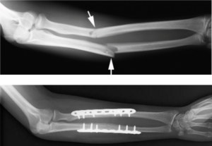
Pre and postoperative xray of forearm fracture.
Surgical Complications
Complications can occur with any surgery, no matter how small. There is always a risk of infection. A dose of antibiotics given prior to surgery helps to make this risk as small as possible. There is always a risk of injury to blood vessels or nerves. This is reduced by having an experienced surgeon involved in your care. It is always possible that the bone may not heal. This is usually associated with patient non-compliance, diabetes, or use of nicotine like smoking and chewing tobacco. If this occurs, secondary surgery may be required. One rare complication of forearm fractures is synostosis. This occurs when excess bone healing occurs between the two bones of the forearm. This results in abnormal stiffness and inability for full motion or rotation. Surgical treatment for this condition is difficult.
Outcomes
Most people with forearm fractures do very well but these fractures are known to be slow to heal sometimes. They take an average of 3-6 months to completely heal. By six weeks, patients are extremely comfortable and usually are released to full activities such as manual labor, skiing and motocross by three to four months. Aggressive return to activity too early can result in re-fracture, hardware breakage or non-union. If you have had surgery, sometimes the metal becomes painful. This can be removed one year after surgery.